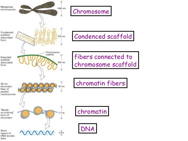

In contrast, micromechanical force measurements suggest that chromosomes have no rigid scaffold 9. Broad axial localization of condensin and Topo IIα have also been observed using EM with immunogold labelling 7, 8. However, due to the diffraction limit of light, the detailed distribution patterns of each scaffold protein are still ambiguous. Immunofluorescence has permitted identification of condensin and Topo IIα within the axial regions of mitotic chromosomes, with their alternating distribution patterns leading to a ‘barber pole’ model 6. Initial electron microscopy (EM) observations of histone-depleted chromosomes suggested that the chromosome scaffold is a network structure 1. Various approaches have been tried to elucidate chromosome scaffold structure. The axially-positioned chromosome scaffold of both chromatids mainly comprises non-histone proteins: so-called scaffold proteins, including condensin, topoisomerase IIα (Topo IIα) and kinesin family member 4 (KIF4) 2, 3, 4, 5. The backbone was positioned along the chromosome axes and thus termed the ‘chromosome scaffold’ 1. In the late 1970s, Laemmli and colleagues observed a backbone structure in chromosomes after depletion of histone proteins. However, how chromosomes are organized in mitosis still remains one of the most important enigmas in cell biology. Our model provides new insights into chromosome higher order structure.Ĭhromosome condensation is crucial to ensure the fidelity of chromosome segregation during cell division. We therefore propose a new structural model of the chromosome scaffold that includes twisted double strands, consistent with the physical properties of chromosomal bending flexibility and rigidity. This reversion to the original morphology underscores the role of the scaffold for intrinsic structural integrity of chromosomes. We also find that scaffold protein can adaptably recover its original localization after chromosome reversion in the presence of cations. Here, we use three dimensional-structured illumination microscopy (3D-SIM) and focused ion beam/scanning electron microscopy (FIB/SEM) to reveal the axial distributions of scaffold proteins in metaphase chromosomes comprising two strands. However, the organization and function of the scaffold are still controversial. The most important structural finding has been the presence of a chromosome scaffold composed of non-histone proteins so-called scaffold proteins. Chromosome higher order structure has been an enigma for over a century.


 0 kommentar(er)
